
How The Ear Works
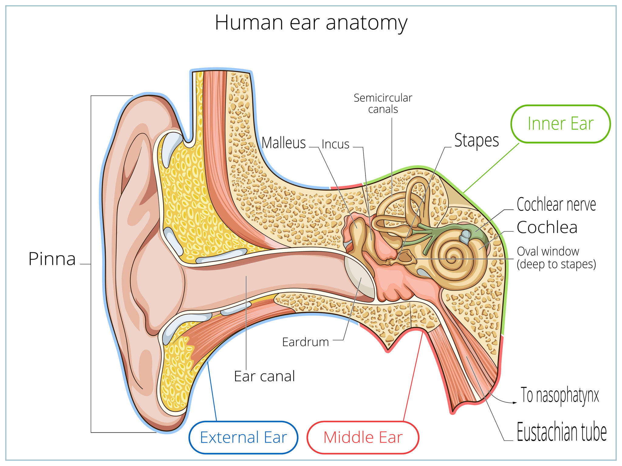
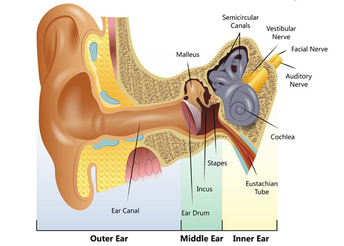
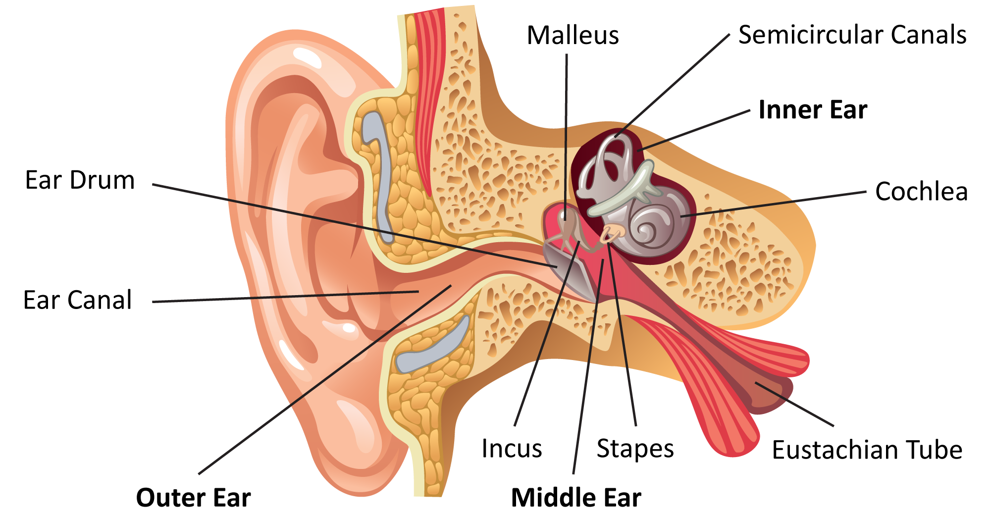
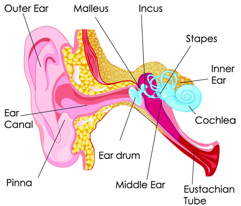
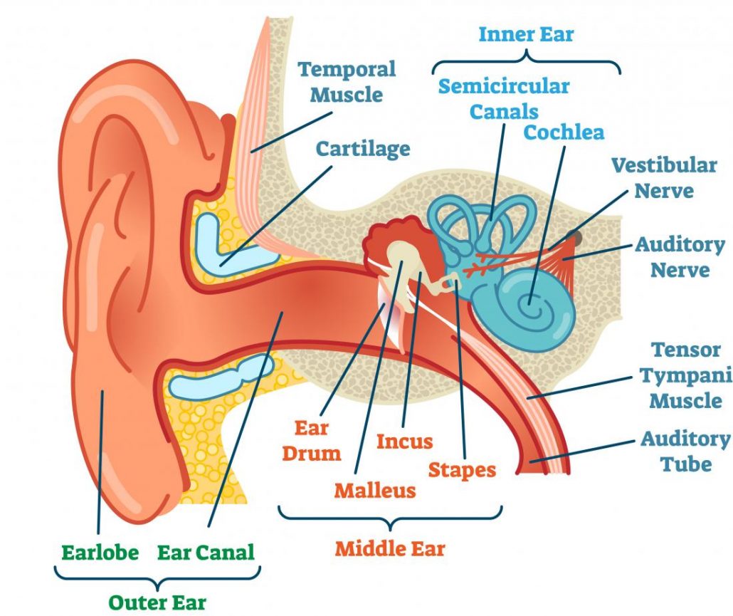
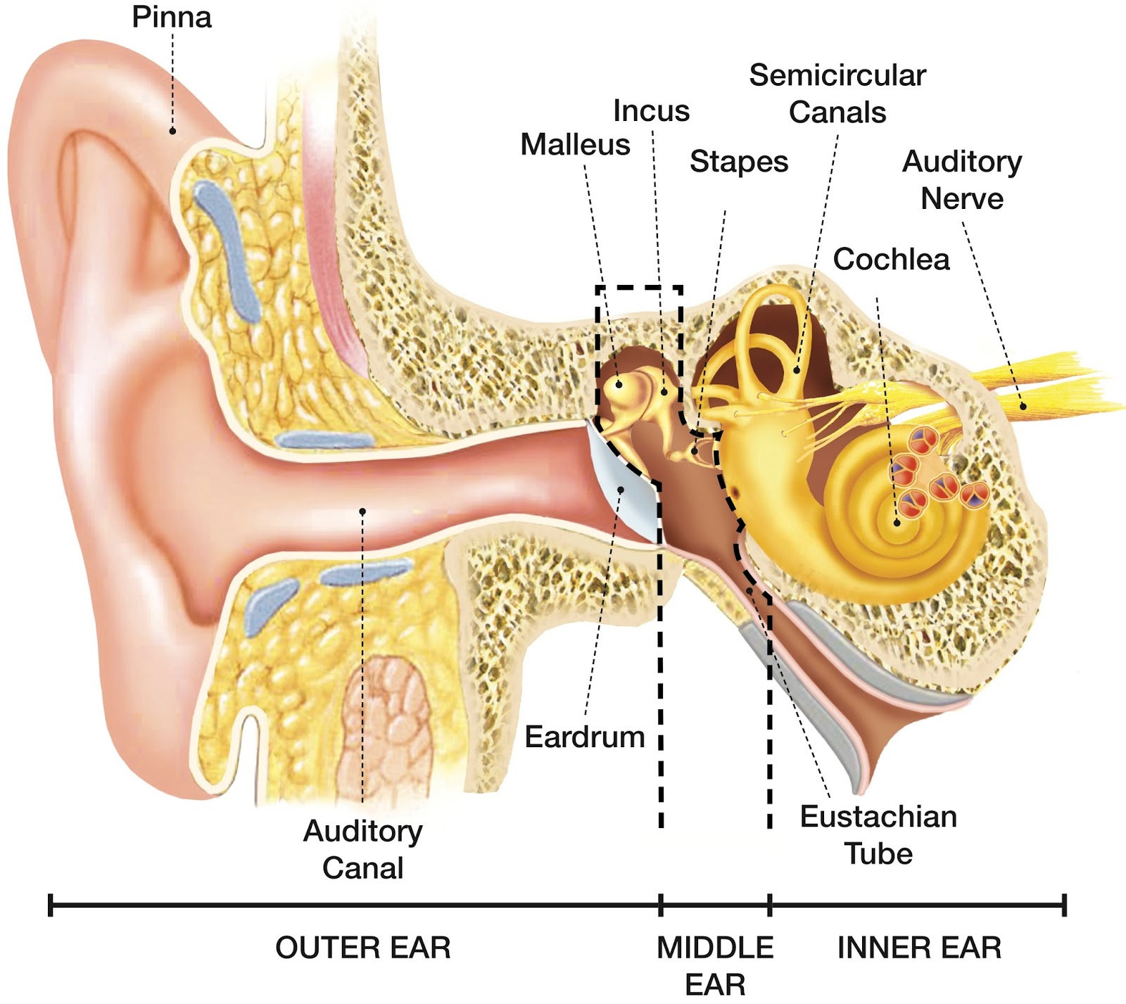
The ear is divided into three parts: Outer ear: The outer ear includes an ear canal that is is lined with hairs and glands that secrete wax. This part of the ear provides protection and.
Ear Anatomy Causes of Hearing Loss Hearing Aids Audiology
Photo name: Ear Diagram Picture category: Human Body Image size: 57 KB Dimensions: 670 x 510 Photo description: This excellent ear diagram labels all the important parts of the human ear system. The labeled parts include the pinna, auditory canal, eardrum, stapes, malleus, incus and cochlea.
Common balance disorders Hearing Link
Anatomy by Shannan Muskopf activity, drag, drop, ear, label Use Google slides to label structures of the ear in this drag and drop activity. Diagram includes the tympanum, ossicles, cochlea, and other organs.

Hearing Health Absolute Hearing
A Diagram of Ear gives us an understanding of the ear's anatomy and its workings. The Human Ear is a sensory organ that processes the sound we hear and passes it to the pinna followed by the transmission of it to the ear canal that results in the vibration of an ear drum.
How We Perceive Sound
1/4 Synonyms: External auditory meatus, External acoustic pore , show more. The ear is a complex part of an even more complex sensory system. It is situated bilaterally on the human skull, at the same level as the nose. The main functions of the ear are, of course, hearing, as well as constantly maintaining balance.

Anatomy of the Ear [4]. Download Scientific Diagram
Your inner ear is the last stop that sound waves make in a carefully orchestrated journey that starts from your outer ear. These waves travel from your outer ear through your middle ear to your inner ear. In the inner ear, the sound waves are converted into electrical energy, which your hearing nerve delivers to your brain as sound, making it.

Hearing Loss Regenerated in Damaged Mammal Ear The Personal Longevity
Label a diagram of the structure of the human ear. Click "Start Assignment". Navigate to the "Science" tab and find the ear diagram. Label the main parts of the ear with Textables and arrows. Add extra information about the functions of the parts of the ear with text boxes.

Alila Medical Media Human ear anatomy, labeled diagram. Medical
Human ear. The ear is divided into three anatomical regions: the external ear, the middle ear, and the internal ear (Figure 2). The external ear is the visible portion of the ear, and it collects and directs sound waves to the eardrum. The middle ear is a chamber located within the petrous portion of the temporal bone.

De anatomie van de oorschelp Health Life Media
Chapter 1 - Introduction Manual Format How to examine the ears Suggested Procedure Chapter 2 - Testing Audiogram Tympanogram Chapter 3 - Ear Anatomy Ear Anatomy - Outer Ear Ear Anatomy - Inner Ear Ear Anatomy Schematics Ear Anatomy Images Chapter 4 - Fluid in the ear Fluid in the ear Discussion Fluid in the ear Outline Middle Ear Ventilation Tubes
The Ear CP Blas de Otero
Your outer ear and middle ear are separated by your eardrum, and your inner ear houses the cochlea, vestibular nerve and semicircular canals (fluid-filled spaces involved in balance and hearing). What is the ear? Your ears are organs that detect and analyze sound. Located on each side of your head, they help with hearing and balance. Advertisement
How noise induced hearing damage and loss occurs
Download a free printable outline of this video and draw along with us: https://artforall.me/video/how-to-draw-human-earThank you for watching. Please subsc.
How The Ear Works Step by Step Brief Explanation
The purpose of the inner ear is to sense and process information about sound and balance, and send that information to the brain. Each part of the inner ear has a specific function. Cochlea: The cochlea is responsible for hearing. It is made up of several layers, with the Organ of Corti at the center.

Strange Ear Comics I Don't Understand
Protect your ears. If the noise is too loud, walk away, turn it down (Turn it to the Left), or use ear plugs. pinna ear canal ear drum hammer anvil stirrup Eustachian tube (connects to the nose) cochlea semicircular canals nerves (connect to the brain) Directions: Color in the diagram below using a different color for each part of the ear.
SPEECH LANGUAGE PATHOLOGY & AUDIOLOGY HEARING DISORDERS OF THE OUTER EAR
Download this blank ear diagram below Contents Ear anatomy overview Ear diagrams (labeled and unlabeled) Accelerate your learning with interactive quizzes Sources + Show all Ear anatomy overview Although it's not obvious to look at, the ear is anatomically divided into three portions: External (outer) ear Middle ear Inner ear

Human Ear Home Tuition Guwahati Assam Human ear diagram, Ear
A brief description of the human ear along with a well-labelled diagram is given below for reference. Well-Labelled Diagram of Ear The External ear or the outer ear consists of Pinna/auricle is the outermost section of the ear. The external auditory canal links the exterior ear to the inner or the middle ear.

labeling the ear Quiz
The ear diagram is one of the important topics for Class 10 and 12 students of the CBSE board and in this article, we will briefly explain the structure of the ear, its different parts and their functions. Parts of the Human Ear. The human ear consists of three different parts. These are: The outer ear. The middle ear. The inner ear
